


Macular Degeneration Treatment
Macular degeneration affects the macula, the small central part of the retina responsible for sharp, straight-ahead sight. It has two forms, dry and wet, and while there is no cure, early detection and disciplined monitoring can slow vision loss. At I Love LASIK and Lens, Samer Khosrof, MD, a fellowship-trained vitreoretinal surgeon, manages the condition with precise imaging and long-term monitoring.
Losing central vision rarely happens all at once. For most patients, macular degeneration starts quietly. There is no pain and no obvious warning. The blur can start in the middle of a page, a face, or a street sign. That gap between when it starts and when you notice it is why early care matters.
At I Love LASIK and Lens, we serve patients across NYC, Manhattan, Brooklyn, and the wider New York area. We combine precise imaging with honest staging. When treatment is needed, options range from nutritional guidance to injectable therapy. Your monitoring plan is built around how quickly your disease is actually progressing, not a standard calendar. Dr. Khosrof is double-trained in vitreoretinal surgery, so the retina is never a secondary consideration here.
- What Is Macular Degeneration?
- What Is the Difference Between Dry and Wet Macular Degeneration?
- What Are the Early Signs of Macular Degeneration?
- How Is Macular Degeneration Diagnosed?
- How Is Macular Degeneration Treated?
- Am I a Candidate for Injectable Treatment?
- Am I at Risk for Macular Degeneration?
- What Can I Do to Protect My Sight?
- Why Do New Yorkers Choose Dr. Khosrof for Macular Degeneration Care?
- FAQs About Macular Degeneration
- Schedule Your Macular Degeneration Consultation

What Is Macular Degeneration?
Macular degeneration is the gradual breakdown of the macula, the small central part of the retina. It is the part that lets you read, recognize faces, and see fine detail directly in front of you. The retina lines the back of the eye and converts light into the signals your brain reads as images. The macula sits at its center. It handles the fine detail: reading a menu, threading a needle, recognizing a face in a crowd.
The disease does not cause total blindness, since peripheral sight generally remains intact. It can, however, quietly hollow out the middle of your visual field. The edges still look normal, which is exactly what makes it easy to overlook. As it progresses, the center of your vision may blur, dim, or develop a blind spot. Your side vision stays comparatively clear.
This is considerably more common than most people expect. A national analysis published in JAMA Ophthalmology found that roughly 19.8 million Americans aged 40 and older have some form of macular degeneration. About 1.49 million of them are in the advanced, sight-threatening stage. The majority sit in the earliest stage and have no symptoms whatsoever.
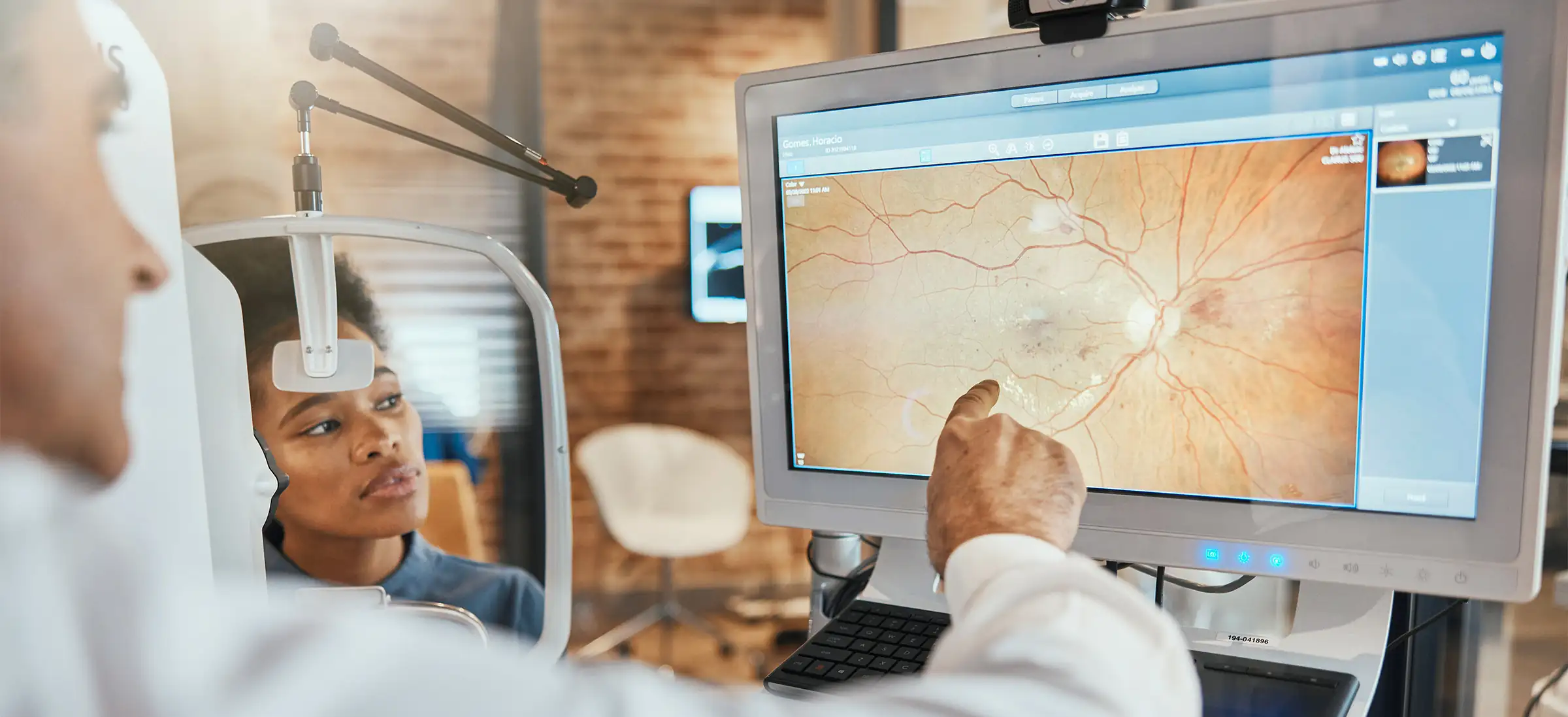
What Is the Difference Between Dry and Wet Macular Degeneration?
Macular degeneration takes one of two forms, and knowing which one you have shapes your entire care plan.
Dry Macular Degeneration
Dry macular degeneration is the far more common form. The macula thins with age, and small yellow deposits of protein and fat, called drusen, build up beneath the retina. It typically advances slowly across years. Its most advanced stage is called geographic atrophy, where patches of retinal tissue die off.
Dr. Khosrof’s own research helped identify one of the biological triggers behind that vessel leakage. That work underlies how today’s injectable treatments target the disease.
Wet Macular Degeneration
Wet macular degeneration is less common but moves fast. Abnormal blood vessels grow beneath the retina and leak fluid or blood into the macula. Vision can change within days rather than years. According to the National Eye Institute, any stage of dry disease can convert to wet. Wet disease is always considered late stage.
These are not two separate diseases. They are two directions the same disease can take. That is why accurate staging matters, and why no single monitoring schedule fits every patient.
What Are the Early Signs of Macular Degeneration?
Early dry macular degeneration usually produces no symptoms at all. That is the single most important thing to understand about it. By the time changes become obvious, structural damage has frequently already occurred.
When symptoms do surface, patients commonly describe:
- Straight lines that appear wavy, bent, or broken
- A blurred, gray, or empty patch at the center of sight
- Needing markedly brighter light to read
- Colors looking duller or washed out
- Trouble recognizing faces before any trouble seeing at distance
The National Eye Institute identifies straight lines appearing wavy as a warning sign of late-stage disease warranting prompt evaluation. If this develops suddenly, treat it as urgent rather than something to raise at your next routine appointment.


How Is Macular Degeneration Diagnosed?
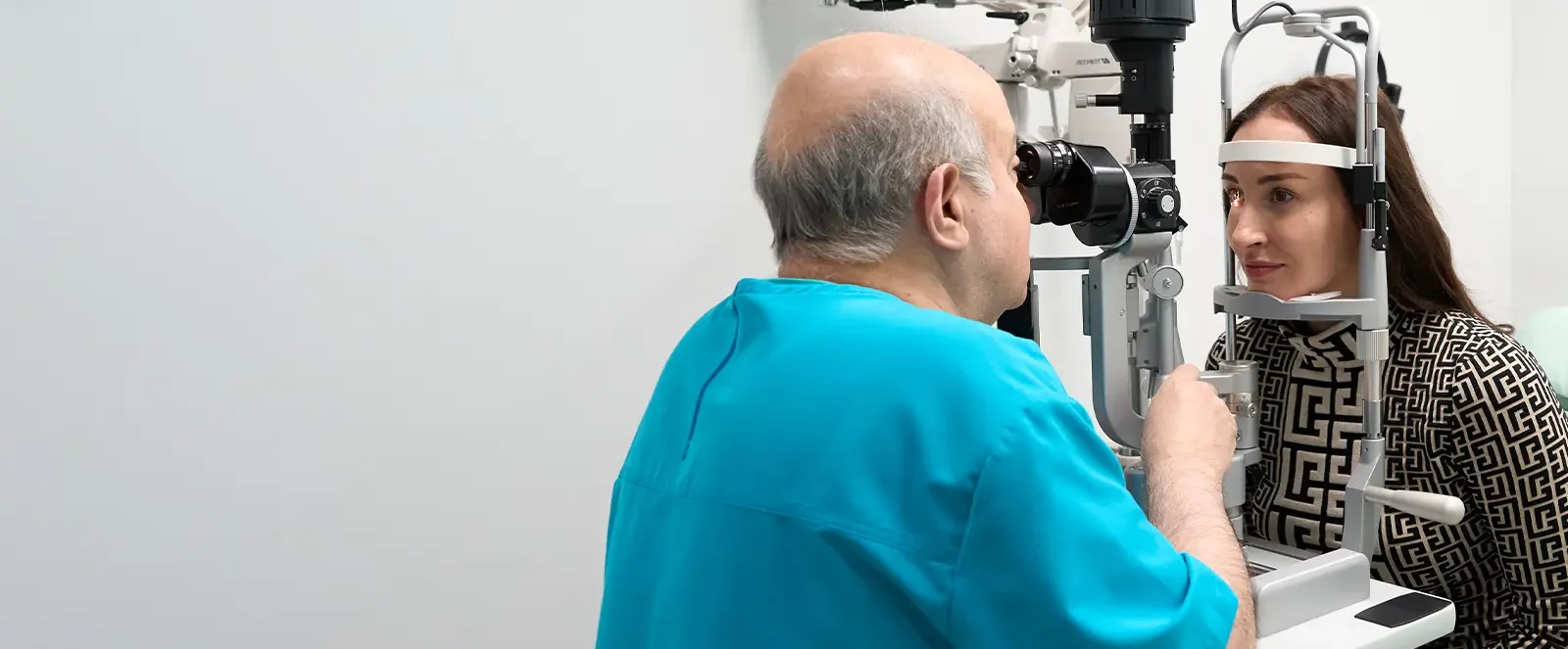
Assessment starts with a dilated retinal exam, which gives your doctor a direct view of the macula. From there, imaging helps confirm the stage of your disease and how it is likely to progress. The main tool is optical coherence tomography, or OCT. It is a quick, painless scan that reveals thinning, fluid, or structural change in the retina. Often, it catches changes before you would notice any difference yourself. In select cases, additional imaging may be used. It gives a closer look at blood flow or leakage beneath the retina.
You will likely also be given an Amsler grid, a simple grid of lines. You check it at home, one eye at a time. It is unglamorous and it works, because you are the person most likely to notice a change first. Reporting distortion between scheduled visits gives your care team the chance to act sooner.

How Is Macular Degeneration Treated?
Treatment depends entirely on which form you have and how far it has advanced.
Treating Dry Macular Degeneration
For intermediate and advanced dry disease, nutritional supplements are the established first step. A Cochrane research review studied the antioxidant and zinc combination used in the Age-Related Eye Disease Study. It found the combination lowered the chance of progressing to late-stage disease. The biggest benefit was for patients who already had intermediate signs. A follow-up trial, AREDS2, found that lutein and zeaxanthin work as a safe substitute for beta-carotene in that formula. This matters because beta-carotene was linked to more lung cancer cases among former smokers. Supplement labels vary widely at the store, so confirm the right formulation with your physician rather than guessing.
Dr. Khosrof’s own research helped show that blocking this pathway meaningfully reduces vessel leakage. That finding helped lay the groundwork for the anti-VEGF treatments used today.
Treating Geographic Atrophy
For geographic atrophy, two injectable medications known as complement inhibitors were approved in 2023. The American Academy of Ophthalmology notes that these medications slowed tissue loss by roughly 14 to 20 percent in trials. They have not been shown to improve eyesight. They also require ongoing injections every one to two months. They are a reasonable option for some patients and not the right choice for others. That decision deserves a real conversation.
Treating Wet Macular Degeneration
For wet disease, anti-VEGF injections placed directly into the eye are the standard of care. They can stabilize or even recover lost vision. Research covering 49 clinical trials and 23,000 eyes found newer medications on extended schedules work about as well as monthly treatment. That means far fewer injections needed over time, a meaningful difference across years of therapy.
Injectable therapy for macular degeneration draws on research Dr. Khosrof contributed to early in his career, and he administers it personally.
Am I a Candidate for Injectable Treatment?
Not everyone with macular degeneration needs injections. We do not proceed unless a patient is confirmed to be a good candidate.
Injectable therapy is indicated for wet disease. The goal is to halt leakage from abnormal vessels and preserve the macula. For geographic atrophy, candidacy is far more individual. The treatment slows tissue loss without restoring sight. The benefit has to be weighed carefully against an indefinite injection schedule and the associated risks. Patients with early or intermediate dry disease are generally better served by supplementation and structured monitoring. This applies when there is no abnormal blood vessel growth.
Determining which category you fall into requires imaging, not an educated guess. That assessment is the purpose of your consultation.
Schedule a Consultation
Am I at Risk for Macular Degeneration?
Age is the biggest factor. CDC data show macular degeneration becomes far more common as you get older. Roughly 2 percent of adults aged 40 to 44 have it, compared with nearly half of adults in the oldest age groups.
Beyond age, a 2025 research review found several risk factors within your control. These include smoking, high blood pressure, heart disease, and diabetes. Of those, smoking showed the strongest link to macular degeneration.
Heredity matters as well. If a parent or sibling carries the diagnosis, your baseline screening should begin earlier than it otherwise would.
What Can I Do to Protect My Sight?
You cannot alter your age or your genetics. But the factors carrying the most weight after those two are within your control. Stopping smoking is the highest-value change available to you. That is because retinal tissue is vascular tissue. Managing blood pressure and blood sugar protects it the same way it protects the kidneys and the heart.
In practice, that means a few habits:
- A diet weighted toward leafy greens, colorful vegetables, and fish
- Sunglasses that block ultraviolet light
- A dilated exam on the schedule your physician sets, not the one your symptoms suggest
If you have been given an Amsler grid, use it. Weekly beats occasionally, and occasionally beats never.
Why Do New Yorkers Choose Dr. Khosrof for Macular Degeneration Care?
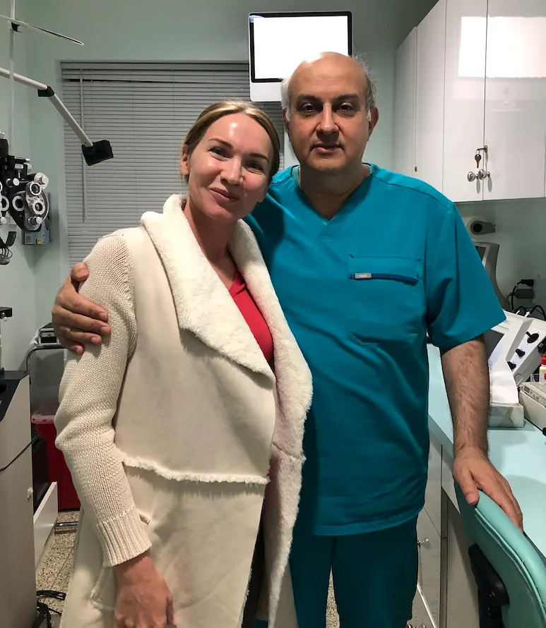
Samer Khosrof, MD, is fellowship-trained in vitreoretinal surgery and double-trained in vitreoretinal surgery, with extensive refractive experience. He has performed thousands of refractive cases alongside his retinal practice. His early research helped shape today’s injectable treatments for macular degeneration, and he administers them personally. We are also among the only practices offering office-based premium cataract surgery and EVO ICL. That matters for macular degeneration patients, who frequently develop cataracts as well and would otherwise be routed between two separate providers.
His engagement with this disease goes beyond the exam room. He has co-authored research on the biological pathway that anti-VEGF injections target in wet macular degeneration today.
What patients tend to remember, though, is the continuity. Dr. Khosrof conducts every consultation, performs every procedure, and handles every follow-up himself, with no handoff to rotating associates. For a condition managed across years and measured in subtle change, that consistency is not a courtesy. It is how deterioration gets caught early. He also contributes his surgical expertise internationally through Physicians Without Borders.
Schedule a Consultation
FAQs About Macular Degeneration
No. The disease affects central sight while sparing peripheral sight. Patients keep the ability to navigate and move around safely even at advanced stages. What it takes first is the detail vision needed for reading, driving, and recognizing faces. Low-vision aids such as magnifiers and adaptive lighting can help preserve independence for daily tasks once central vision is affected.
Within days, not weeks. Sudden distortion can signal conversion from dry to wet disease. Same-week evaluation gives the best chance to start treatment before scarring develops. Call the office directly rather than waiting for your next scheduled visit. An urgent visit typically includes updated imaging the same day, so treatment can begin quickly if needed.
Most patients begin with a loading phase of monthly injections. After that, the interval extends according to how the retina responds on imaging. Some stabilize at longer intervals on newer agents, per the extended-dosing research cited above. Others continue to need more frequent treatment. Your schedule is set by your scans, not by a fixed protocol.
Yes, family history raises your risk. If you have been diagnosed, share that with your adult siblings and children. That way, they can start dilated screening earlier than the general population would.
Cost depends on which form of the disease you have, what imaging is required, and whether injectable therapy is indicated. Because of that, it cannot be quoted meaningfully in advance. Our team reviews the full financial picture with you before any treatment begins. That includes anything payable at the time of your visit.
Often yes. Cataract and macular degeneration affect entirely different structures, so removing a clouded lens can still improve functional sight. Your ceiling, however, is set by the health of the macula, so expectations must be established candidly beforehand. Dr. Khosrof evaluates both conditions in the same appointment rather than splitting them across two practices.

Schedule Your Macular Degeneration Consultation
You may have noticed distortion, or a dim or absent spot at the center of your sight. Or you may carry a family history and want a baseline on record. Either way, the next step is a dilated retinal evaluation with imaging. We serve patients from across NYC, Manhattan, Brooklyn, and greater New York, and Dr. Khosrof conducts every consultation personally.