

Diagnosis and Treatment of Retinal Disorders
Retinal disorders include retinal detachment, retinal vein occlusion, floaters and diabetic retinopathy, and they range from harmless to sight-threatening. Some need attention within hours, while others are watched carefully over years. At I Love LASIK and Lens, Samer Khosrof, MD, is fellowship-trained in vitreoretinal surgery and personally evaluates, treats and follows each retinal patient. A dilated retinal examination is the only dependable way to know which situation you are in.
Retinal disorders are conditions that affect the retina, the thin layer of light-sensitive tissue lining the back of the eye. Because the retina turns light into the signals your brain reads as vision, damage there affects sight directly and sometimes permanently. Patients across NYC, Manhattan, Brooklyn and the wider New York area come to I Love LASIK and Lens for retinal care that stays with a single surgeon from the first examination through recovery. Dr. Khosrof manages consultation, treatment and follow-up himself, so the person who finds the problem is the person who treats it and tracks how your eye responds.
- What Are Retinal Disorders?
- What Causes a Retinal Detachment, and Why Is It Urgent?
- What Is Retinal Vein Occlusion, and How Is It Treated?
- When Are Floaters Harmless, and When Are They a Warning Sign?
- How Does Diabetic Retinopathy Damage Vision?
- How Are Retinal Disorders Diagnosed?
- Am I a Good Candidate for Retinal Treatment or Surgery?
- Why Should I Choose Dr. Samer Khosrof for Retinal Care?
- What Do Patients Say About Their Retinal Care?
- What Else Do Patients Ask About Retinal Disorders?
- How Do I Schedule a Consultation?

What Are Retinal Disorders?
The retina lines the inside of the eye the way film lines the back of a camera. When it tears, loses blood flow, swells or pulls loose, vision changes in ways glasses cannot correct. Four presentations account for most retinal visits. A detachment lifts the retina off the eye wall. A vein occlusion blocks drainage from part of the retina and causes swelling. Floaters are drifting shapes caused by changes in the gel that fills the eye. Diabetic retinopathy damages the small retinal blood vessels over years of elevated blood sugar. These conditions share an organ and little else, which is why each calls for a different examination and a different plan.
What Causes a Retinal Detachment, and Why Is It Urgent?
Most detachments begin with a small tear. As the gel inside the eye shrinks with age, it can tug hard enough to open a break in the retina, and fluid then seeps behind the tissue and separates it from its blood supply. Nearsightedness, prior eye surgery and eye injury all raise the risk. The warning signs are specific: a sudden shower of new floaters, flashes of light, or a dark curtain moving in from the side of your vision. None of it hurts, which is exactly why people wait.
Waiting costs vision. Once the retina loses its blood supply, the light-sensing cells begin to fail, and the longer central vision stays detached, the less of it returns. Repair may involve laser or freezing treatment, a gas bubble, a supporting band placed outside the eye, or vitreoretinal surgery to remove the gel from inside the eye. Results are good but rarely instant. In a published series of adults aged 18 to 40, the retina was reattached in roughly two thirds of eyes with one operation and in more than 97 percent after further surgery, which is why we discuss the possibility of a second procedure before the first one.
Schedule a Consultation
What Is Retinal Vein Occlusion, and How Is It Treated?
A retinal vein occlusion is a blocked drainage vein in the retina. Pressure builds behind the blockage, fluid leaks into the tissue, and vision blurs or dims, often noticed on waking. Pooled population data put the prevalence at about 5.2 cases per 1,000 adults aged 30 and older, with branch occlusions far more common than central ones, and an estimated 16 million adults affected worldwide.
Because a blocked retinal vein is a vascular event, the finding matters beyond the eye. Blood pressure, cholesterol, blood sugar and clotting all belong in the conversation, and Dr. Khosrof coordinates with your physician when the picture suggests uncontrolled systemic disease. Treatment targets the swelling rather than the clot. Injections that block vascular endothelial growth factor are the accepted first-line therapy for the resulting central swelling, and a portion of patients who respond poorly do better after a switch to a steroid-based option. Injections are given in the office, and the schedule is usually heavier at first and lighter once the retina stabilizes.
When Are Floaters Harmless, and When Are They a Warning Sign?
Floaters are shadows cast by strands in the vitreous gel, and most are a normal part of aging. The distinction that matters is timing. Floaters you have lived with for years are rarely a threat. Floaters that appear suddenly, multiply quickly or arrive with flashes deserve a dilated examination promptly, because that pattern can accompany a retinal tear.
An initially clean examination is reassuring but not final. In one long-term study of patients with acute floaters and flashes and no tear at diagnosis, about 7 percent developed a delayed tear over roughly six years, with most of those tears appearing within the first year. That is why we schedule a follow-up rather than simply sending you home. For long-standing floaters that genuinely interfere with reading or driving, surgical removal of the gel is possible, and reported patient satisfaction is high, though there are real risks. Dr. Khosrof treats that operation as a considered decision, not a routine one.
How Does Diabetic Retinopathy Damage Vision?
Diabetes injures the smallest blood vessels first, and the retina is full of them. Vessels weaken, leak and close off, and the eye responds by growing fragile new vessels that bleed and form scar tissue capable of pulling the retina loose. Among people living with diabetes, roughly 22 percent have some diabetic retinopathy and about 6 percent have the vision-threatening form. Swelling of the central retina, which affects about one in 15 people with diabetes, is the most common reason vision drops.
The early stages produce no symptoms at all, so how well you see is not a useful gauge of whether damage has started. Treatment depends on stage. Injections that block vascular endothelial growth factors can slow and in some cases reverse the disease, and laser treatment remains valuable in advanced cases. In a randomized trial of proliferative disease, 97 percent of patients treated with injections retained 20/40 vision or better in at least one eye at two years, compared with 87 percent treated with scatter laser. When bleeding fills the eye or scar tissue detaches the retina, surgery is the answer.
How Are Retinal Disorders Diagnosed?
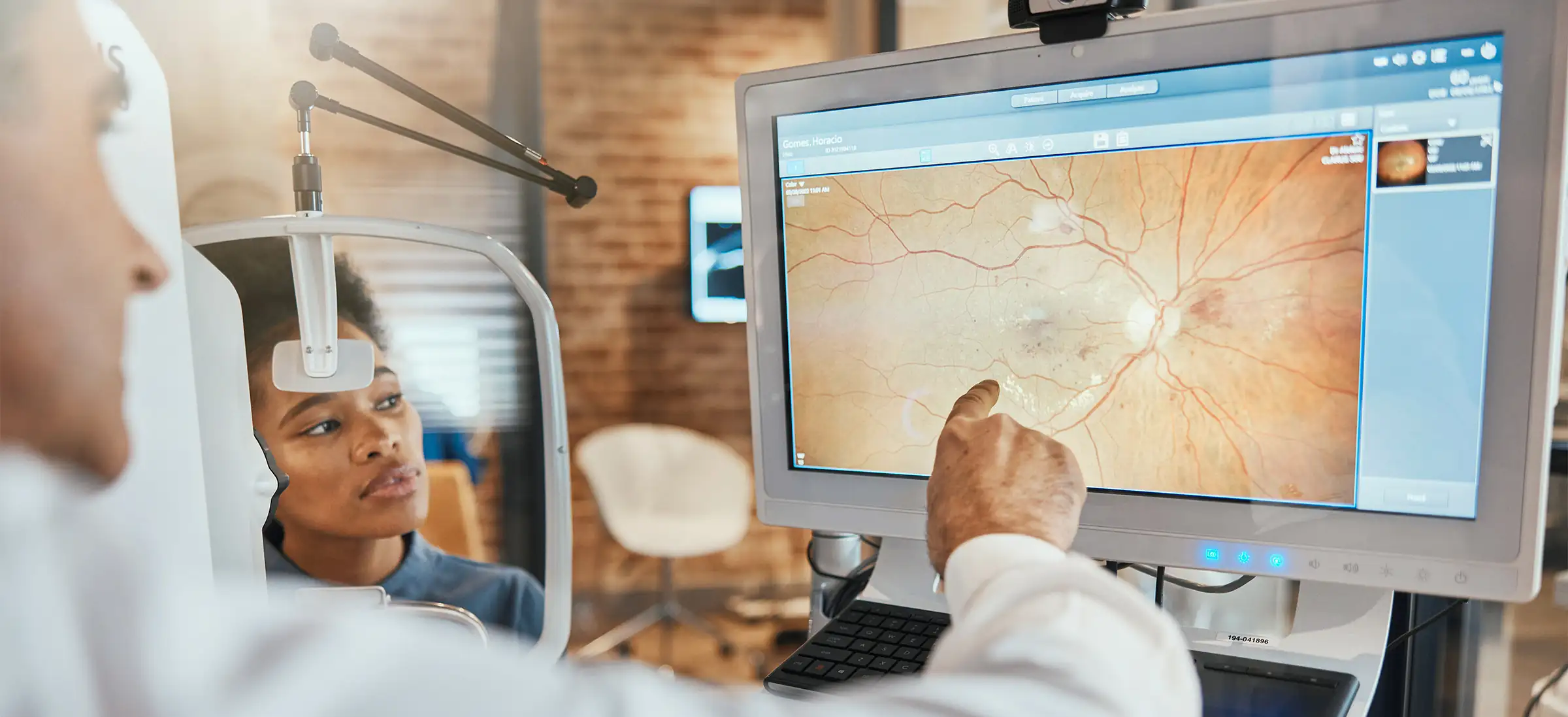
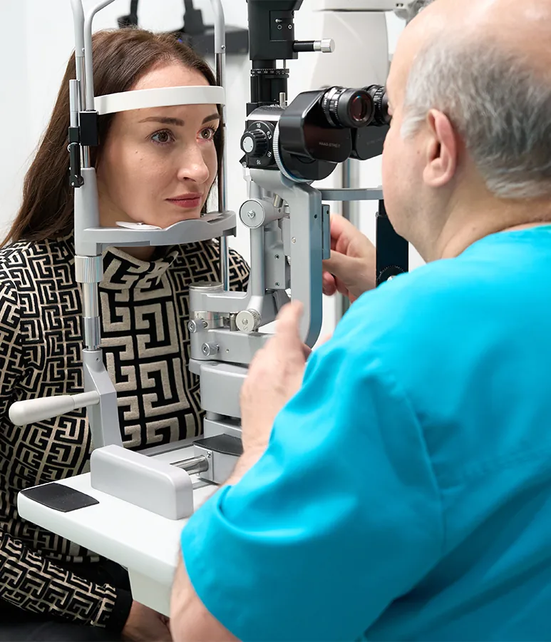
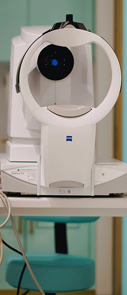
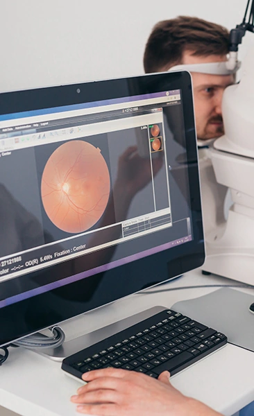
Diagnosis starts with dilating drops and a careful look at the full retina, including the far edges where tears hide. Imaging fills in what the examination cannot show. Optical coherence tomography maps the layers of the central retina and measures swelling to the micron. Angiography traces blood flow and reveals leaking or closed vessels. Ultrasound helps when blood inside the eye blocks the view. Dr. Khosrof reviews these studies himself and explains what he is seeing while you are still in the chair, so the plan is understood before you leave.
Am I a Good Candidate for Retinal Treatment or Surgery?
Candidacy depends on the condition, the anatomy and the timing. A fresh retinal tear with good central vision is usually an excellent candidate for prompt sealing. A detachment involving the center of vision still warrants surgery, though expectations must be managed about how much sight returns. Swelling from vein occlusion or diabetes responds best when treated before the central retina has thinned and scarred.
Some eyes are not good candidates for a given procedure. Long-standing detachments with heavy scar tissue, eyes with no remaining light perception, and floaters that are visually trivial all belong in a different conversation. Uncontrolled blood sugar or blood pressure may need attention before elective retinal surgery. Dr. Khosrof does not proceed unless the examination confirms you are a suitable candidate, and if a different approach or a different specialist serves you better, you will be told so plainly.
Schedule a Consultation
Why Should I Choose Dr. Samer Khosrof for Retinal Care?
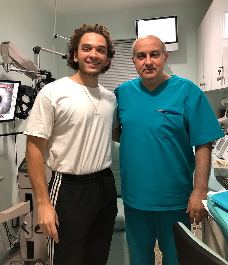
Samer Khosrof, MD, is fellowship-trained in vitreoretinal surgery, the subspecialty devoted to the retina and the gel in front of it. He is double-trained, and his research contributed to the development of the modern injectable treatments now used for macular degeneration.
What patients notice is the continuity. Dr. Khosrof runs a single-surgeon practice by design: he performs your examination, he performs your procedure, and he sees you at every post-operative visit. Nothing is handed to a rotating team, and no one has to be brought up to speed on your eye. He is one of only a few physicians to offer office-based surgery for select lens and refractive procedures, including EVO ICL and premium cataract procedures, and he performs humanitarian eye surgery with Physicians Without Borders.
What Do Patients Say About Their Retinal Care?

Doctor very nice and staff very polite. Doctor took very good care. Recommend all my friends.D.S.
I love this doc . I think he is the best eye doc :)M.E.
He is very thorough and talks you throughout your procedure. His assistant is equally wonderful. Very caring staff and can't say enough about the doctor.E.H.

Schedule a Consultation
What Else Do Patients Ask About Retinal Disorders?
Recovery ranges from a few days to several weeks depending on the procedure. Laser sealing of a tear involves almost no downtime. Gas bubble repair requires holding a specific head position for days and rules out air travel until the bubble absorbs. Vitrectomy and scleral buckle recovery generally spans several weeks, with vision improving gradually over months rather than immediately.
Yes, though late recurrence after a fully successful repair is uncommon. A large surgical review identified 39 late recurrences among 16,396 operations. Diabetic retinopathy and vein occlusion behave differently, since both are ongoing conditions that require continued monitoring even after vision recovers.
The National Eye Institute advises a dilated eye exam at least once a year for everyone with diabetes. If retinopathy is already present, exams may be needed every two to four months. Pregnancy raises the risk, so a dilated exam should be scheduled as early in pregnancy as possible.
Consultations at I Love LASIK and Lens are a paid appointment rather than a free screening. Because detachment, vein occlusion and diabetic retinopathy are medical conditions rather than elective procedures, coverage differs from coverage for elective vision correction, and specific benefits should be verified with your plan.
Not necessarily, but plan on a course rather than a single treatment. Injections are typically given more often during the first year and less often once the retina stabilizes. Some patients taper off entirely, others need occasional maintenance, and the interval is set by what imaging shows at each visit.

Ready to Schedule a Consultation?
If your vision has changed suddenly, if floaters or flashes have appeared, or if you have diabetes and have not had a dilated exam this year, schedule a consultation with Dr. Khosrof. I Love LASIK and Lens serves patients throughout NYC, Manhattan, Brooklyn and New York, and retinal symptoms are treated as time-sensitive.